
Living with persistent pain, numbness, or tingling can significantly impact your daily life. Understanding the source of these symptoms is the first step toward finding relief. Nerve compression, also known as a pinched nerve, occurs when surrounding tissues, bones, or disc material apply pressure to a nerve. This restricts the nerve’s ability to function properly and causes a range of uncomfortable symptoms. For many patients in the Greater Dallas area, patients nerve compression symptoms often develop gradually due to degenerative changes, repetitive activities, or injury. Recognizing these symptoms early and seeking evaluation from a board-certified spine specialist can prevent progression and help you return to the activities you enjoy.
Understanding Nerve Compression
Nerve compression develops when pressure is applied to a nerve by surrounding structures in the spine or elsewhere in the body. This pressure interferes with the nerve’s normal signal transmission, leading to pain, numbness, weakness, and other symptoms. These symptoms can range from mildly annoying to debilitating.
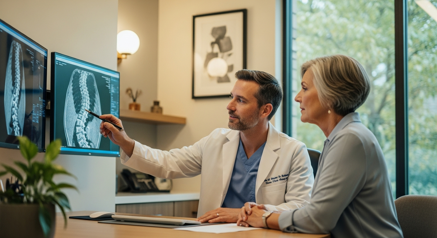
The spinal nerves are particularly vulnerable to compression because they must travel through narrow passages formed by bones, discs, and ligaments. often The cervical spine (neck), thoracic spine (mid-back), and lumbar spine (lower back) are common sites where nerve compression occurs. When a nerve becomes compressed, it cannot effectively transmit signals between your brain and the parts of your body it serves. This is why symptoms often appear in areas distant from the actual compression site.
The importance of early diagnosis and treatment cannot be overstated. potentially causing irreversible weakness, loss of sensation, or chronic pain. What begins as occasional discomfort can progress to constant symptoms that interfere with work, sleep, and quality of life. A comprehensive evaluation by a fellowship-trained spine specialist can identify the source of compression and guide you toward the most appropriate treatment options.
Common Types of Nerve Compression Syndromes
Different types of nerve compression syndromes affect various areas of the body. Each has characteristic symptoms and patterns. Understanding these distinctions helps patients recognize their condition and communicate effectively with their healthcare providers.
Cervical radiculopathy occurs when a nerve in the neck is compressed, often causing pain that radiates down the arm. This condition frequently results from disc herniation or bone spurs in the cervical spine. Patients often describe sharp, burning pain accompanied by numbness or tingling following the affected nerve’s pathway.
Lumbar radiculopathy involves nerve compression in the lower back and commonly causes sciatica. Pain radiates from the lower back through the buttock and down the leg. may The sciatic nerve is the largest nerve in the body, which explains why sciatica symptoms can be particularly intense and disruptive to daily activities.
Carpal tunnel syndrome affects the median nerve at the wrist. It is characterized by numbness and tingling in the thumb and first three fingers. While not directly related to the spine, this condition shares similar mechanisms of nerve compression. It often affects Dallas-area professionals usually who spend extended hours at computer workstations.
Piriformis syndrome develops when the piriformis muscle in the buttock compresses the sciatic nerve. This creates symptoms similar to lumbar radiculopathy but arising from a different anatomical location. Distinguishing between these conditions requires expert evaluation and diagnostic testing.
Symptoms and Warning Signs
Recognizing the symptoms of nerve compression enables patients to seek timely evaluation and treatment. While symptoms vary depending on which nerve is affected, certain patterns are common across different types of nerve compression.
Nerve compression often causes radiating pain that travels along the nerve pathway from the compression site. This radiating quality distinguishes nerve pain from localized muscle soreness or joint pain. Many patients describe the sensation as sharp, shooting, or electric in quality.
Numbness and tingling in the hands, feet, or limbs are common indicators of nerve compression. These sensations may be constant or intermittent. They often worsen with certain positions or activities that increase pressure on the affected nerve.
Weakness in the affected areas represents a more serious symptom that warrants prompt attention. Progressive weakness that interferes with daily activities suggests the need for professional evaluation by a spine specialist. If you notice difficulty holding objects, walking, or performing tasks that were previously easy, contact a spine care provider without delay.
What Causes Nerve Compression
Understanding the underlying causes of nerve compression helps patients comprehend their condition and make informed decisions about treatment. Multiple factors can contribute to nerve compression, often working in combination.
Herniated discs are a leading cause of nerve compression. They occur when the soft inner material of a disc protrudes outward, potentially pressing against nearby nerves. The intervertebral discs serve as cushions between vertebrae. When they herniate, the displaced material can occupy space normally reserved for nerve roots.
Bone spurs develop as part of osteoarthritis and can narrow the space available for nerves in the spine. These bony growths represent the body’s response to degenerative changes. Unfortunately, they can create new problems by encroaching on nerve pathways.
typically Ligament thickening, particularly of the ligamentum flavum in the spinal canal, reduces the available space for nerves and the spinal cord. This thickening often develops gradually with age and repetitive stress on the spine.
Lifestyle and occupational factors play significant roles in nerve compression development. Repetitive activities and poor posture can contribute to nerve compression over time, particularly in patients in the Dallas area who work sedentary jobs. Extended periods in fixed positions increase pressure on spinal structures and can accelerate degenerative processes.
Risk Factors
Certain populations face elevated risk for developing nerve compression. Awareness of these risk factors can promote preventive measures and early intervention.
Age is a significant risk factor. usually Degenerative changes in the spine become more common after age 50. While younger patients can certainly experience nerve compression from injury or other causes, the incidence increases substantially with advancing age.
Occupations requiring repetitive motions or prolonged sitting increase the risk of nerve compression. For many professionals in the Greater Dallas area, understanding occupational risk factors enables implementation of preventive ergonomic strategies.
Previous spine injuries or trauma can accelerate degenerative processes that lead to nerve compression. Even injuries that occurred years earlier can influence current spinal health and nerve compression risk.
Genetic predisposition also contributes to nerve compression susceptibility. Some families demonstrate higher rates of degenerative disc disease and spinal arthritis. While you cannot control your genetic inheritance, awareness enables proactive management through lifestyle modifications and early intervention when symptoms develop.
Diagnosis: How Spine Specialists Evaluate Nerve Compression
Accurate diagnosis forms the foundation of effective treatment. Modern spine care utilizes multiple diagnostic tools to pinpoint the exact source and severity of nerve compression.
The diagnostic process begins with a comprehensive patient history and physical examination. Your spine specialist will ask detailed questions about symptom onset, location, quality, and aggravating factors. The physical examination includes neurological testing to assess strength, sensation, and reflexes in the affected areas. This helps identify which specific nerve is compressed.
MRI imaging is the gold standard for diagnosing nerve compression. It provides detailed views of soft tissues and nerve compression sites. Unlike X-rays, which show only bones, MRI scans visualize the spinal cord, nerve roots, discs, and surrounding soft tissues with exceptional clarity.
CT scans may be utilized when MRI is not suitable or when detailed bone imaging is needed. X-rays help evaluate alignment, bone spurs, and structural stability, though they cannot directly visualize nerve compression.
Electromyography (EMG) and nerve conduction studies (NCS) measure how well nerves transmit electrical signals and can confirm nerve damage. These electrodiagnostic tests provide functional information about nerve health that complements the anatomical information from imaging studies.
A board-certified spine specialist performs a comprehensive evaluation combining imaging, physical examination, and patient history to develop an accurate diagnosis. This thorough approach distinguishes expert spine care from more generic pain management.
Treatment Options: From Conservative to Surgical Care
Treatment for nerve compression follows a graduated approach. It begins with conservative management and progresses to more invasive interventions only when necessary. This patient-centered strategy respects your body’s natural healing capacity while ensuring access to advanced care when required.
Conservative treatment, including rest, physical therapy, and anti-inflammatory medications, successfully resolves symptoms in many patients with nerve compression. Your spine specialist may recommend activity modification, avoiding positions or movements that aggravate symptoms while maintaining overall fitness through low-impact activities.
Physical therapy and rehabilitation play central roles in conservative management. A structured therapy program addresses muscle imbalances, improves flexibility, and strengthens supporting structures to reduce pressure on compressed nerves. Therapists may also employ manual techniques, traction, and modalities to promote healing and symptom relief.
Medications including nonsteroidal anti-inflammatory drugs (NSAIDs), muscle relaxants, and sometimes neuropathic pain medications can help manage symptoms during the healing process. These medications address inflammation, muscle spasm, and nerve pain through different mechanisms. Your physician will select appropriate options based on your specific presentation.
Epidural steroid injections can provide temporary relief by reducing inflammation around compressed nerves. These injections deliver anti-inflammatory medication directly to the affected area. They often provide weeks to months of symptom improvement while other treatments take effect.
Surgical intervention is considered when conservative treatments fail to provide relief after 6-12 weeks or when nerve damage is progressive. The decision to proceed with surgery represents a collaborative process between you and your spine surgeon, weighing symptom severity, functional limitations, and treatment goals.
Minimally invasive procedures require smaller incisions than traditional open surgery. This leads to less tissue damage and faster recovery times. Fellowship-trained surgeons at Legent Spine utilize advanced minimally invasive techniques to achieve excellent outcomes while minimizing surgical trauma.
Minimally Invasive Surgical Approaches
When conservative treatments have been exhausted and surgical intervention becomes necessary, minimally invasive approaches offer significant advantages over traditional open procedures. These advanced techniques enable spine surgeons to address nerve compression effectively while minimizing collateral tissue damage.
Microdiscectomy is a minimally invasive procedure that removes the herniated portion of a disc to relieve nerve compression. This procedure is highly effective for disc-related radiculopathy. Many patients experience rapid symptom improvement following surgery.
Laminoforaminotomy enlarges the opening where the nerve exits the spine, reducing compression and associated pain. This decompression procedure addresses stenosis without requiring fusion in many cases, preserving normal spinal motion.
Endoscopic procedures utilize a tiny camera and instruments inserted through minimal incisions. This allows surgeons to visualize and treat nerve compression with exceptional precision. These techniques result in minimal muscle disruption and often allow same-day discharge.
Minimally invasive approaches typically result in shorter hospital stays, reduced blood loss, and faster return to normal activities compared to open surgery. The reduced tissue trauma translates to less postoperative pain and quicker recovery, enabling earlier return to work and daily activities.
Recovery and What to Expect After Treatment
Understanding the recovery process helps patients set realistic expectations and actively participate in their healing journey. Recovery timelines vary depending on treatment type, severity of nerve compression, and individual factors. However, general patterns apply to most patients.
Patients undergoing minimally invasive procedures often experience symptom improvement within 2-4 weeks. While pain relief may occur rapidly, complete nerve healing requires more time. Damaged nerves regenerate slowly over weeks to months.
Structured physical therapy is essential after treatment to rebuild strength and prevent recurrence. Your therapist will guide you through progressive exercises designed to restore normal movement patterns, strengthen supporting muscles, and improve flexibility without overstressing healing tissues.
Activity restrictions vary based on the specific procedure performed and your individual healing progress. Most patients can perform light daily activities soon after minimally invasive surgery, with gradual increases in activity as healing progresses. Your surgeon will provide specific guidelines regarding lifting restrictions, return to work, and resumption of exercise or sports.
Full recovery and return to normal activities typically takes 6-8 weeks for minimally invasive procedures, compared to 12+ weeks for open surgery. Patients who actively participate in rehabilitation and follow postoperative guidelines generally achieve better outcomes than those who remain passive in their recovery.
Prevention: Protecting Your Spine Health
While not all nerve compression is preventable, many risk factors can be modified through conscious lifestyle choices and healthy habits. Implementing preventive strategies reduces your risk of developing nerve compression and supports overall spinal health.
Regular core strengthening exercises can reduce the risk of nerve compression by providing better spinal stability. Strong abdominal and back muscles act as a natural brace for your spine, distributing forces more evenly during daily activities.
Proper ergonomics and regular breaks during prolonged sitting significantly lower the risk of developing nerve compression, especially for Dallas-area office workers who spend extended hours at computer workstations. Adjusting your workspace to support neutral spine positions, using ergonomic furniture, and taking brief movement breaks every 30-60 minutes all contribute to spine health.
Regular exercise promotes circulation, maintains disc nutrition, and prevents the muscle weakness and stiffness that contribute to nerve compression. Activities like walking, swimming, and yoga provide excellent whole-body conditioning without excessive spinal stress.
Maintaining a healthy body weight reduces stress on the spine and associated structures that can cause nerve compression. Each pound of body weight translates to multiple pounds of force on spinal structures during movement and activity.
Proper lifting techniques protect your spine from acute injury and cumulative damage. Bending at the knees rather than the waist, holding objects close to your body, and avoiding twisting while lifting all reduce injury risk. For Greater Dallas residents, these principles apply whether lifting boxes at work, gardening at home, or playing with grandchildren.
Expert Nerve Compression Care in the Greater Dallas Area
When nerve compression symptoms interfere with your daily life, accessing expert spine care close to home makes all the difference. Legent Spine provides world-class spine care to patients throughout the Greater Dallas area with board-certified, fellowship-trained specialists.
Our spine surgeons, including Dr. Paul Salinas and Dr. Grant Booher, have completed advanced fellowship training in minimally invasive spine surgery. They bring the latest techniques and technologies to patients in Dallas and surrounding communities. This specialized training ensures access to the most advanced treatment options without traveling to distant medical centers.
The practice utilizes advanced minimally invasive techniques to treat nerve compression with smaller incisions and faster recovery times. From initial evaluation through recovery and beyond, Legent Spine maintains a commitment to transparent, honest communication and personalized care.
Legent Spine’s dedicated care coordinators work with each patient to develop personalized treatment plans that address their unique condition and goals. This team-based approach combines surgical expertise with attentive support, creating an environment where patients feel heard, respected, and confident in their care.
If you’re experiencing symptoms of nerve compression, seeking evaluation from a spine specialist represents an important step toward relief and recovery. Early intervention prevents progression and expands treatment options, giving you the best opportunity for complete symptom resolution. Contact Legent Spine to schedule a comprehensive evaluation and begin your journey toward better spine health and improved quality of life.